Bacillus velezensis VM54: A UV-tolerant Strain with Antifungal Activity against Diverse Phytopathogens
DOI:
https://doi.org/10.29356/jmcs.v70i1.2610Keywords:
Antifungal, chromium-tolerant strain, fengycin, MALDI-TOF MS, surfactin, tomatoAbstract
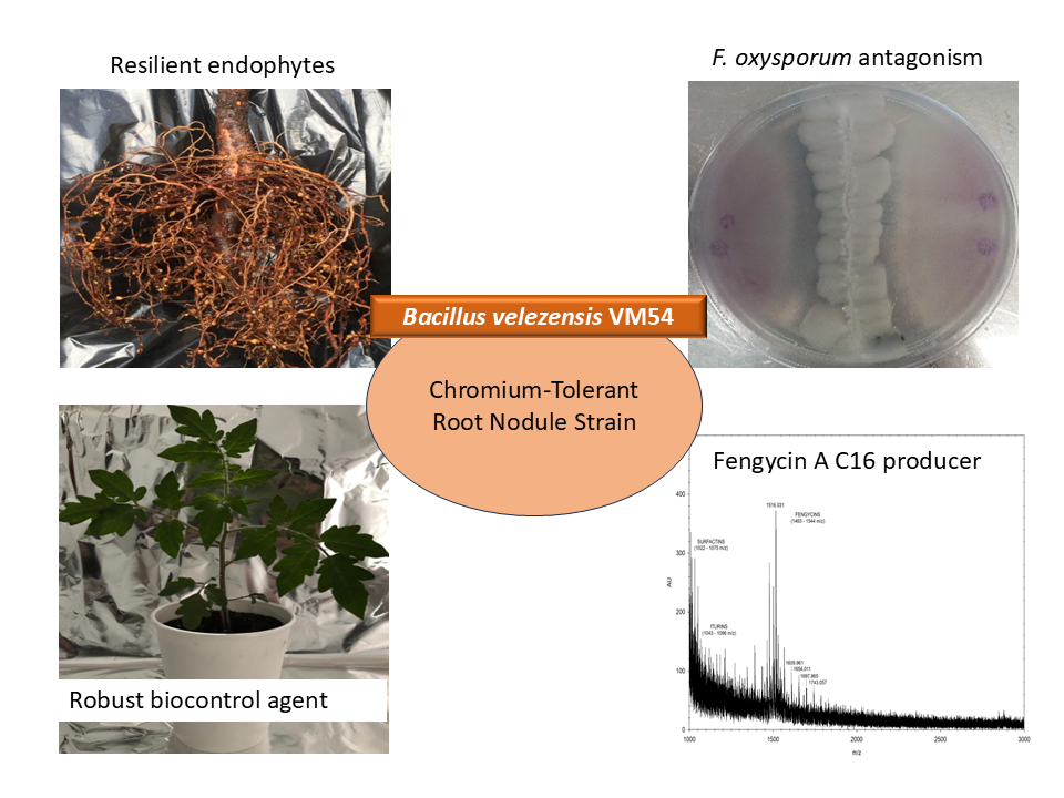
Fungal diseases significantly affect crop yield and product quality. The use of chemical fungicides to control fungal infestations can disturb the plant-associated microbiome and raise concerns about food security. Therefore, the use of biological antifungals, such as Bacillus species, is encouraged as they offer a sustainable alternative to chemical fungicides. However, the success of such biocontrol agents in the field depends on their ability to withstand environmental stressors like solar UV light and dryness. Here, we present a targeted strategy for selecting resilient biocontrol agents by first isolating endophytic microorganisms that are already adapted to extreme conditions, followed by screening them for antifungal activity. Using this approach, we investigated the chromium-tolerant, root nodule-associated strain Bacillus velezensis VM54 for its ability to antagonize fungal pathogens. B. velezensis VM54 exhibited strong in vitro and in vivo antagonism against Fusarium fujikuroi, F. oxysporum, and Alternaria alternata. Notably, the VM54 strain tolerated UV-C radiation up to 1824 J/m², a level comparable to that of the extremophile Deinococcus radiodurans, highlighting its robustness for potential field application. PCR-based genome analysis revealed the presence of biosynthetic gene clusters for several antimicrobial compounds, including surfactin, bacillaene, difficidin, and macrolactins. Mass spectrometry of cell-free culture extracts confirmed the production of putative fengycin A C16 and C17 lipopeptides.
These bioactive metabolites, along with volatile compounds produced by VM54, are likely responsible for its antifungal activity. This study supports the use of environmentally adapted endophytes as a promising source of resilient biocontrol agents for sustainable agriculture.
Resumen. Las enfermedades causadas por hongos afectan significativamente el rendimiento de los cultivos y la calidad del producto. El uso de fungicidas químicos para controlar las infestaciones fúngicas puede alterar el microbioma asociado a las plantas y generar riesgos para la seguridad alimentaria. Por lo tanto, el uso de antifúngicos biológicos es altamente recomendable, ya que ofrece una alternativa sostenible a los fungicidas químicos. Sin embargo, el éxito de estos agentes de biocontrol en el campo depende de su capacidad para resistir estresores ambientales como la luz solar UV y la sequía. En este trabajo, presentamos una estrategia para seleccionar agentes de biocontrol resilientes mediante el aislamiento de microorganismos endófitos ya adaptados a condiciones extremas, seguida de un escrutinio de la actividad antifúngica. Con este enfoque, investigamos la cepa Bacillus velezensis VM54, tolerante al cromo y asociada a nódulos radiculares, debido a su capacidad de antagonizar patógenos fúngicos. B. velezensis VM54 exhibió un fuerte antagonismo in vitro e in vivo frente a Fusarium fujikuroi, F. oxysporum y Alternaria alternata. Cabe destacar que la cepa VM54 toleró la radiación UV-C hasta 1824 J/m², un nivel comparable al de extremófilo Deinococcus radiodurans, lo que evidencia su robustez para su posible aplicación en el campo. El análisis genómico basado en ensayos de PCR reveló la presencia de grupos de genes biosintéticos para varios compuestos antimicrobianos, incluidos surfactina, bacillaeno, difficidina y macrolactinas. La espectrometría de masas de los extractos de cultivos confirmó la producción de los supuestos lipopéptidos C16 y C17 de fengicina A. Estos metabolitos bioactivos, junto con los compuestos volátiles producidos por VM54, probablemente son los responsables de la actividad antifúngica. Este estudio respalda el uso de endófitos adaptados al medio ambiente como una fuente prometedora de agentes de biocontrol resilientes para la agricultura sostenible.
Downloads
References
1. Chen, X.; Huang, H.; Zhang, S.; Zhang, Y.; Jiang, J.; Qiu, Y.; Liu, J.; Wang, A. Agriculture. 2021, 11, 581. DOI: https://doi.org/10.3390/agriculture11070581
2. Kai, K.; Wang, R.; Bi, W.; Ma, Z.; Shi, W.; Ye, Y.; Zhang, D. World J. Microbiol. Biotechnol. 2021, 37,93. DOI: https://doi.org/10.1007/s11274-021-03062-x
3. Niehaus, E.M.; Kim, H.K.; Münsterkötter, M.; Janevska, S.; Arndt, B.; Kalinina, S.A.; Houterman, P.M.; Ahn, P.M.; Alberti, I.; Tonti, S.; Kim, D.W.; Sieber, C.M.K.; Humpf, H.U.; Yun S.H.; Güldener, U.; Tudzynski, B. PLOS Pathogens. 2017, 13, e1006670. DOI: https://doi.org/10.1371/journal.ppat.1006670
4. Karačić, V.; Miljaković, D.; Marinković, J.; Ignjatov, M.; Milošević, D.; Tamindžić, G.; Ivanović, M. Microorganisms. 2024, 12, 457. DOI: https://doi.org/10.3390/microorganisms12030457
5. Abdelaziz, A.M.; Elrefaey, A.A.; Sharaf, M.H.; Al‑Qthanin, R.N.; Attia, M.S. Sci. Rep. 2024, 14, 18052. DOI: https://doi.org/10.1038/s41598-024-67233-0
6. Moghaddam, G.A.; Rezayatmand, Z.; Nasr Esfahani, M.; Khozaei, M. Plant Physiol. Biochem. 2019, 142, 500-509. DOI: https://doi.org/10.1016/j.plaphy.2019.08.011
7. Gardener, B.B.; Fravel, D.R. Plant Health Progress. 2002, 3, 17. DOI: https://doi.org/10.1094/PHP-2002-0510-01-RV
8. Hazarika, D.J.; Goswami, G.; Gautom, T.; Parveen, A.; Das, P.; Barooah, M.; Boro, R.C. BMC Microbiol. 2019, 19, 71. DOI: https://doi.org/10.1186/s12866-019-1440-8
9. Mateus, J.R.; Dal’Rio, I.; Jurelevicius, D.; Faria da Mota, F.; Marques, J.M.; Thiago Juca Ramos, R.; da Costa da Silva, A.L.; Gagliardi, P.R.; Seldin, L. Agriculture. 2021, 11, 1046. DOI: https://doi.org/10.3390/agriculture11111046
10. Rabbee, M.F.; Hwang, B.S.; Baek, K.H. Agronomy. 2023, 13, 840. DOI: https://doi.org/10.3390/agronomy13030840
11. 11. Idris, A.L.; Li, W.; Huang, F.; Lin, F.; Guan, X.; Huang, T. World J. Microbiol. Biotechnol. 2024, 40, 58. DOI: https://doi.org/10.1007/s11274-023-03856-1
12. Ramírez, V.; Baez, A.; López, P.; Bustillos, R.; Villalobos, M.A.; Carreño, R.; Contreras, J.L.; Muñoz-Rojas, J.; Fuentes, L.E.; Martínez, J.; Munive, J.A. Front. Microbiol. 2019, 10,1833. DOI: https://doi.org/10.3389/fmicb.2019.01833
13. Ramirez, V.; Martínez, J.; Bustillos‐Cristales, M.R.; Castañeda‐Antonio, D.; Munive, J.A.; Baez, A. J. Appl. Microbiol. 2022, 132, 470-482. DOI: https://doi.org/10.1111/jam.15179
14. Martens, M.; Dawyndt, P.; Coopman, R.; Gillis, M.; De Vos, P.; Willems, A. Int. J. Syst. Evol. Microbiol. 2008, 58, 200-14. DOI: https://doi.org/10.1099/ijs.0.65392-0
15. Thompson, J.D.; Gibson, T.J.; Plewniak, F.; Jeanmougin, F.; Higgins, D.G. Nucleic Acids Res. 1997, 25, 4876-82. DOI: https://doi.org/:10.1093/nar/25.24.4876
16. Nicholas, K.B.; Nicholas, J.H. Berlin: Science Open, Inc. 1997.
17. Rzhetsky, A.; Nei, M. Mol. Biol. Evol. 1992, 9, 945-967. DOI: https://doi.org/10.1093/oxfordjournals.molbev.a040771
18. Tamura, K.; Stecher, G.; Kumar, S. Mol. Biol. Evol. 2021, 38, 3022-3027. DOI: https://doi.org/10.1093/molbev/msab120
19. Figueiredo JEF, Marins MS, Silva FC, Ribeiro VP, Diniz GDFD, Rodrigues VA, Oliveira-Paiva CA, Lana UGDP, Ferreira LVS, Sousa Tinoco SMD, Cruz-Magalhães V. Microbiol. Resour Announc. 14, e01279-24. DOI: https://doi.org/10.1128/mra.01279-24
20. Bodour, A.A.; Miller-Maier, R.M. J. Microbiol. Methods. 1998, 32, 273-280. DOI: https://doi.org/10.1016/S0167-7012(98)00031-1
21. Corral-Lugo, A.; Morales-García, Y.E.; Pazos-Rojas, L.A.; Ramírez-Valverde, A.; Martínez-Contreras, R.D.; Muñoz-Rojas, J. Rev. Colomb. Biotecnol. 2012, 14, 147-156.
22. Haddad, N.I.; Wang, J.; Mu, B. Protein Pept. Lett. 2009, 16, 7-13. DOI: https://doi.org/10.2174/092986609787049358
23. Zhao, X.; Begyn, K.; Delongie, Y.; Rajkovic, A.; Uyttendaele, M. Food Microbiol. 2023, 115, 104325. DOI: https://doi.org/10.1016/j.fm.2023.104325
24. Bauermeister, A.; Bentchikou, E.; Moeller, R.; Rettberg, P. Arch. Microbiol. 2009, 191, 913-8. DOI: https://doi.org/10.1007/s00203-009-0522-7
25. Bruno, L.B.; Karthik, C.; Ma, Y.; Kadirvelu, K.; Freitas, H.; Rajkumar, M. Chemosphere. 2020, 244, 125521. DOI: https://doi.org/10.1016/j.chemosphere.2019.125521
26. Din, B.U.; Amna Rafique, M.; Javed, M.T.; Kamran, M.A.; Mehmood, S.; Khan, M.; Sultan, T.; Hussain Munis, M.F.; Chaudhary, H.J. Plant. Physiol. Biochem. 2020, 146, 249-258. DOI: https://doi.org/10.1016/j.plaphy.2019.11.010
27. Matilda, C.S.; Mannully, S.T.; Viditha, R.P.; Shanthi, C. J. Appl. Microbiol. 2019, 127, 121-133. DOI: https://doi.org/10.1111/jam.14293
28. Shreya, D.; Jinal, H.N.; Kartik, V.P.; Amaresan, N. Arch. Microbiol. 2020, 202, 887-894. DOI: https://doi.org/:10.1007/s00203-019-01801-1
29. Lin, L.Z.; Zheng, Q.W.; Wei, T.; Zhang, Z.Q.; Zhao, C.F.; Zhong, H.; Xu, Q.Y.; Lin, J.F.; Guo, L.Q. Front. Microbiol. 2020, 11, 579621. DOI: https://doi.org/10.3389/fmicb.2020.579621
30. Bai, X.; Li, Q.; Zhang, D.; Zhao, Y.; Zhao, D.; Pan, Y.; Wang, J.; Yang, Z.; Zhu, J. Biology (Basel). 2013, 12, 856. DOI: https://doi.org/10.3390/biology12060856
Downloads
Published
Issue
Section
License
Copyright (c) 2026 Verónica Ramírez, Angelina Palomino-García, Emmanuel Anguiano-García, Daniel Balleza, José-Antonio Munive, Antonino Baez

This work is licensed under a Creative Commons Attribution-NonCommercial 4.0 International License.
Authors who publish with this journal agree to the following terms:
- Authors retain copyright and grant the journal right of first publication with the work simultaneously licensed under a Creative Commons Attribution License that allows others to share the work with an acknowledgement of the work's authorship and initial publication in this journal.
- Authors are able to enter into separate, additional contractual arrangements for the non-exclusive distribution of the journal's published version of the work (e.g., post it to an institutional repository or publish it in a book), with an acknowledgement of its initial publication in this journal.