Size-Specific Synthesis and Biological Evaluation of Silver Nanoparticles: A Computational and Experimental Integration
DOI:
https://doi.org/10.29356/jmcs.v70i1.2410Keywords:
silver nanoparticles, nanomaterials, design of experiments, cytotoxicity, cell viabilityAbstract
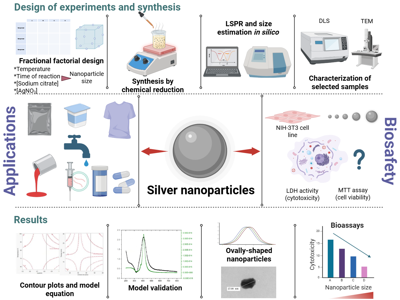
During the last years, nanomaterials, such as silver nanoparticles (AgNPs), have revolutionized various areas due to their antimicrobial properties. However, their impact on human health in the short, medium, and long term has yet to be fully understood due to the variability in their sizes and the lack of standards that define a specific size and their biological impact. Specialized software can help develop mathematical models and predict the conditions necessary to produce AgNPs of a controlled size. These tools complement experimental techniques, facilitating physical-chemical characterization and contributing to a more precise regulation and safe use of AgNPs in various applications. This study aimed to determine the optimal conditions for the chemical synthesis of AgNPs of different sizes through the design of experiments (DOE) to optimize the synthesis conditions and to evaluate their effects in the NIH-3T3 cell line. A 24 DOE was carried out, varying the temperature, reaction time, concentration of the precursor agent, and concentration of the reducing agent, using the nanoparticle size as a response variable supported by the MiePlot software. AgNPs were characterized by ultraviolet-visible light absorption spectroscopy, dynamic light scattering, and transmission electron microscopy. It was possible to synthesize and characterize the AgNPs with a predominant size of 60 nm, which conditions were also complemented with the MiePlot software. It was found that temperature and the concentration of reducing agents influence nanoparticle size and that the smaller the nanoparticle, the more significant toxicity they exhibit in NIH-3T3 cells. The present study shows the value of the complementary use of computational tools like MiePlot integrated with experimental methods for the size-specific synthesis of AgNPs. These findings provide a reference point for comparing and predicting the biological effects of similarly sized AgNPs, offering a broader framework for their safe and controlled application in various fields.
Resumen. Durante los últimos años, los nanomateriales, como las nanopartículas de plata (AgNPs), han revolucionado diversas áreas debido a sus propiedades antimicrobianas. Sin embargo, su impacto en la salud humana a corto, mediano y largo plazo aún no se comprende completamente debido a la variabilidad en sus tamaños y la falta de estándares que definan un tamaño específico y su impacto biológico. El software especializado puede ayudar a desarrollar modelos matemáticos y predecir las condiciones necesarias para producir AgNPs de un tamaño controlado. Estas herramientas complementan las técnicas experimentales, facilitando la caracterización físico-química y contribuyendo a una regulación más precisa y al uso seguro de las AgNPs en diversas aplicaciones. Este estudio tuvo como objetivo determinar las condiciones óptimas para la síntesis química de AgNPs de diferentes tamaños mediante el diseño de experimentos (DOE) para optimizar las condiciones de síntesis y evaluar sus efectos en la línea celular NIH-3T3. Se llevó a cabo un DOE 24, variando la temperatura, el tiempo de reacción, la concentración del agente precursor y la concentración del agente reductor, utilizando el tamaño de la nanopartícula como variable de respuesta apoyada por el software MiePlot. Las AgNPs se caracterizaron mediante espectroscopía de absorción de luz ultravioleta-visible, dispersión de luz dinámica y microscopía electrónica de transmisión. Fue posible sintetizar y caracterizar las AgNPs con un tamaño predominante de aproximadamente 60 nm, cuyas condiciones también se complementaron con el software MiePlot. Se encontró que la temperatura y la concentración de agentes reductores influyen en el tamaño de las nanopartículas y que cuanto más pequeña es la nanopartícula, mayor toxicidad exhiben en las células NIH-3T3. El presente estudio muestra el valor del uso complementario de herramientas computacionales como MiePlot integradas con métodos experimentales para la síntesis específica de AgNPs por tamaño. Estos hallazgos proporcionan un punto de referencia para comparar y predecir los efectos biológicos de AgNPs de tamaño similar, ofreciendo un marco más amplio para su aplicación segura y controlada en diversos campos.
Downloads
References
1. McGillicuddy, E.; Murray, I.; Kavanagh, S.; Morrison, L.; Fogarty, A.; Cormican, M.; Dockery, P.; Prendergast, M.; Rowan, N.; Morris, D. Sci. Total. Environ. 2017, 575, 231-246. DOI: https://doi.org/10.1016/J.SCITOTENV.2016.10.041
2. Islam, M. A.; Jacob, M. V.; Antunes, E. J. Environ. Manage. 2021, 281, 111918. DOI: https://doi.org/10.1016/J.JENVMAN.2020.111918
3. Natsuki, J.; Natsuki, T.; Hashimoto, Y. Int. J. Mater. Sci. 2015, 4, 325–32. DOI: https://doi.org/10.11648/J.IJMSA.20150405.17
4. Syafiuddin, A.; Salmiati; Salim, M. R.; Beng Hong Kueh, A.; Hadibarata, T.; Nur, H. Chin. J. Chem. 2017, 64, 732–56. DOI: https://doi.org/10.1002/JCCS.201700067
5. Sánchez Moreno, M. Máster Universitario en Ciencia y Tecnología Química. Universidad Nacional de Educación a Distancia, 2017.
6. Lee, S. H.; Jun, B. H. Int. J. Mol. Sci. 2019, 20, 865. DOI: https://doi.org/10.3390/IJMS20040865
7. Li, X.; Wang, L.; Fan, Y.; Feng, Q.; Cui, F. Z. J. Nanomater. 2012, 54838. DOI: https://doi.org/10.1155/2012/548389
8. Haider, A.; Kang, I. K. Adv. Mat. Sci. Eng. 2015, 1–16. DOI: https://doi.org/10.1155/2015/165257
9. Augustine, R.; Hasan, A.; Primavera, R.; Wilson, R.J.; Thakor, A.S.; Kevadiya, B. D. Mater. Today Commun. 2020, 25, 101692. DOI: https://doi.org/10.1016/j.mtcomm.2020.101692
10. Rohde, M. M.; Snyder, C.M.; Sloop, J.; Solst, S. R.; Donati, G. L.; Spitz, D. R.; Furdui, C. M.; Singh, R. Part. Fibre Toxicol. 2021, 18, 37. DOI: https://doi.org/10.1186/s12989-021-00430-1
11. Ferdous, Z.; Nemmar, A. Int. J. Mol. Sci. 2020, 21, 2375. DOI: https://doi.org/10.3390/ijms21072375
12. Prabhu, S.; Poulose, E. K. Int. Nano Lett. 2012, 2, 1–10. DOI: https://doi.org/10.1186/2228-5326-2-32
13. Pulit-Prociak, J.; Stokłosa, K.; Banach, M. Environ. Chem. Lett. 2015, 13, 59–68. DOI: https://doi.org/10.1007/S10311-014-0490-2/TABLES/1
14. Montgomery, D. C., in: Design and Analysis of Experiments. 10th ed., Wiley, Hoboken, 2020.
15. https://www.mt.com/us/en/home/library/white-papers/automated-reactors/Innovative-Techniques-to-Synthesize-Breakthrough-Molecule.html, accessed in October 2024.
16. Montoya-Marquez, J.; Sánchez-Estudillo, L.; Torres-Hernández, P. Ciencia y Mar. 2011, 15, 61–70.
17. http://www.philiplaven.com/mieplot.htm, accessed in February 2024.
18. Barbir, D.; Dabic, P.; Mehes, M. Hem. Ind. 2019, 73, 397–404. DOI: https://doi.org/10.2298/HEMIND190719031B
19. Gakiya-Teruya, M.; Palomino-Marcelo, L.; Rodriguez-Reyes, J. C. F. Methods Protoc. 2019, 2, 3. DOI: https://doi.org/10.3390/MPS2010003
20. Torres, Y.; López, I.; Balderas, I.; Arredondo, E.; González, P.; Ramírez, M. Revista de Ciencias Farmacéuticas y Biomedicina. 2017, 1, 19.
21. Dong, X.; Ji, X.; Wu, H., Zhao, L.; Li, J.; Yang, W. J. Phys. Chem. C. 2009, 113, 6573–6. DOI: https://doi.org/10.1021/jp900775b
22. Khodashenas, B.; Ghorbani, H. R. Arab. J. Chem. 2019, 12, 1823–38. DOI: https://doi.org/10.1016/J.ARABJC.2014.12.014
23. Yerragopu, P. S.; Hiregoudar, S.; Nidoni, U.; Ramappa, K. T.; Sreenivas, A. G.; Doddagoudar, S. R. Int. Res. J. Pure Appl. Chem. 2020, 21, 37-50. DOI: https://doi.org/10.9734/IRJPAC/2020/V21I330159
24. Quintero-Quiroz, C.; Acevedo, N.; Zapata-Giraldo, J.; Botero, L. E.; Quintero, J.; Zárate-Triviño, D.; Saldarriega, J.; Pérez, V. Z. Biomater. Res. 2019, 23. DOI: https://doi.org/10.1186/s40824-019-0173-y
25. Colón Figuerona, M. Ingeniería en Materiales. Benemérita Universidad Autónoma de Puebla, 2015.
26. Bohren, C. F.; Huffman, D. R., in: Absorption and Scattering of Light by Small Particles. WILEY‐VCH Verlag GmbH & Co. KGaA. Weinheim, 1998. DOI: https://doi.org/10.1002/9783527618156
27. Danaei, M.; Dehghankhold, M.; Ataei, S.; Hasanzadeh Davarani, F.; Javanmard, R.; Dokhani, A.; Khoeasani, S.; Mozafari, M. R. Pharmaceutics. 2018, 10, 57. DOI: https://doi.org/10.3390/pharmaceutics10020057
28. Bhattacharjee, S. J. Control Release. 2016, 235, 337–51. DOI: https://doi.org/10.1016/J.JCONREL.2016.06.017
29. Kaushik, R.; Saran, S.; Isar, J.; Saxena, R. K. J. Mol. Catal. B Enzym. 2006, 40, 121–6. DOI: https://doi.org/10.1016/J.MOLCATB.2006.02.019
30. Jaramillo, M.; Ossa-Orozco, C. P. Revista ION. 2020, 33, 17–32. DOI: https://doi.org/10.18273/revion.v33n1-2020002
31. Schluesener, J. K.; Schluesener, H. J. Arch. Toxicol. 2013, 87, 569–76. DOI: https://doi.org/10.1007/S00204-012-1007-Z/FIGURES/3
32. María, D.; Delgadillo Álvarez, C. Revista Fesahancccal. 2021, 7, 17–24.
33. Jiravova, J.; Tomankova, K. B.; Harvanova, M.; Malina, L.; Malohlava, J.; Luhova, L.; Panacek, A.; Manisova, B.; Kolarova, H. Food Chem. Toxicol. 2016, 96, 50–61. DOI: https://doi.org/10.1016/J.FCT.2016.07.015
34. Chueh, P. J.; Liang, R. Y.; Lee, Y. H.; Zeng, Z. M.; Chuang, S. M. J. Hazard Mater. 2014, 264, 303–12. DOI: https://doi.org/10.1016/j.jhazmat.2013.11.031
35. Amooaghaie, R.; Saeri, M. R.; Azizi, M. Ecotoxicol. Environ. Saf. 2015, 120, 400–8. DOI: https://doi.org/10.1016/j.ecoenv.2015.06.025
36. Barbasz, A.; Oćwieja, M.; Roman, M. Colloids Surf. B Biointerfaces. 2017, 156, 397–404. DOI: https://doi.org/10.1016/j.colsurfb.2017.05.027
37. Bélteky, P.; Rónavári, A.; Zakupszky, D.; Boka, E.; Igaz, N.; Szerencsés, B.; Kiricsi, M.; Kónya, Z. Int. J. Nanomedicine. 2021, 16, 3021–40. DOI: https://doi.org/10.2147/IJN.S304138
Downloads
Published
Issue
Section
License
Copyright (c) 2026 Aída J. Velarde-Salcedo, Karen O Ruiz-Reyes, Gabriela Navarro-Tovar, Carmen Gonzalez

This work is licensed under a Creative Commons Attribution-NonCommercial 4.0 International License.
Authors who publish with this journal agree to the following terms:
- Authors retain copyright and grant the journal right of first publication with the work simultaneously licensed under a Creative Commons Attribution License that allows others to share the work with an acknowledgement of the work's authorship and initial publication in this journal.
- Authors are able to enter into separate, additional contractual arrangements for the non-exclusive distribution of the journal's published version of the work (e.g., post it to an institutional repository or publish it in a book), with an acknowledgement of its initial publication in this journal.