Effects of Firing on Iron content of Wyoming Montmorillonite: Mössbauer and Photoacoustic Study
DOI:
https://doi.org/10.29356/jmcs.v70i1.2520Keywords:
Firing, Montmorillonite, Structural iron, Mössbauer, Isomer shift, Quadrupole splittingAbstract
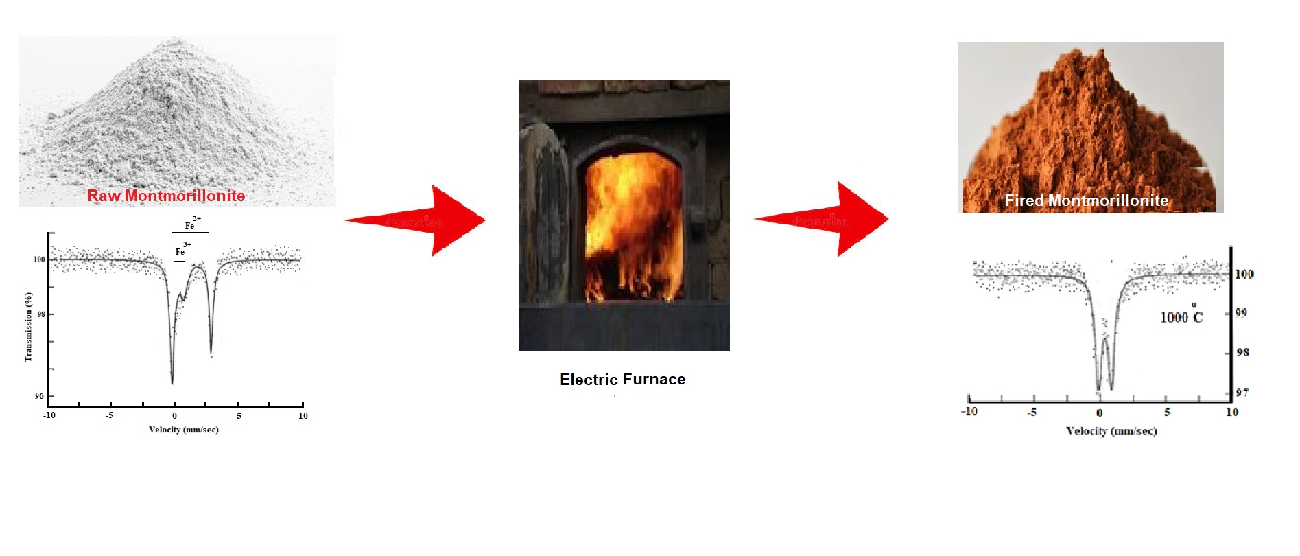
Montmorillonite MMT is a naturally occurring aluminosilicate layered mineral that is made of AlO6 (octahedron) and SiO4 (tetrahedron), sharing top oxygen atoms. The iron oxidation state in the crystal lattice of MMT clay is critical in determining the surface and colloidal chemistry. The changes in the behavior of the structural iron in Wyoming MMT clay, upon firing from 100 oC up to 1000 oC were investigated. The transformation of iron was monitored using Mössbauer spectroscopy as the main tool, and other changes were studied using photoacoustic spectroscopy. The clay samples were fired from 100 up to 1000 oC. No formal change in the Mössbauer parameters was observed on firing the sample from 100 to 500 oC except the oxidation of Fe(II) at about 200 oC. The main changes in the Fe(III) Mössbauer parameters occurred upon firing from 600 oC up to 1000 oC. The isomer shift decreases gradually with increasing temperature of firing, due to the de-hydroxylation of the structure. The quadrupole splitting of the Fe(III) doublet increased, from 600 oC to 1000 oC, indicating an increase in distortion of the lattice as the temperature was raised. A slight increase in the linewidth of Fe(III) values of the fired samples from that of the original sample was also observed, suggesting that more than one iron site occurs. The photoacoustic investigations were in good agreement with Mössbauer results.
Resumen. La montmorillonita MMT es un mineral aluminosilicato laminar de origen natural, constituido por unidades de AlO₆ (octaedros) y SiO₄ (tetraedros), que comparten los átomos de oxígeno de sus vértices. El estado de oxidación del hierro en la red cristalina de la arcilla MMT es un factor crítico para determinar su química superficial y coloidal. Se investigaron los cambios en el comportamiento del hierro estructural presente en la montmorillonita de Wyoming al someterla a calcinación entre 100 °C y 1000 °C. La transformación del hierro se monitoreó principalmente mediante espectroscopía Mössbauer, mientras que otros cambios se estudiaron utilizando espectroscopía fotoacústica. Las muestras de arcilla fueron calcinadas a temperaturas comprendidas entre 100 y 1000 °C. No se observaron cambios significativos en los parámetros Mössbauer al calcinar las muestras entre 100 y 500 °C, con excepción de la oxidación del Fe(II), que ocurrió alrededor de 200 °C. Los principales cambios en los parámetros Mössbauer correspondientes al Fe(III) se produjeron al incrementar la temperatura de calcinación de 600 a 1000 °C. El desplazamiento isomérico disminuyó gradualmente con el aumento de la temperatura de calcinación, como consecuencia de la deshidroxilación de la estructura. El desdoblamiento cuadrupolar del doblete de Fe(III) aumentó entre 600 y 1000 °C, indicando un incremento en la distorsión de la red cristalina a medida que se elevaba la temperatura. Asimismo, se observó un ligero aumento en el ancho de línea de las señales de Fe(III) de las muestras calcinadas con respecto a la muestra original, lo que sugiere la existencia de más de un sitio cristalográfico para el hierro. Los estudios fotoacústicos mostraron una excelente concordancia con los resultados obtenidos mediante espectroscopía Mössbauer.
Downloads
References
1. Rathossi, C.; Pontikes, Y. J. Eur. Ceram. Soc. 2010, 30, 1841-51. DOI: https://doi.org/10.1016/j.jeurceramsoc.2010.02.002.
2. Pontikes, Y.; Angelopoulos, G. N. Adv. Appl. Ceram. 2009, 108, 50-6. DOI: https://doi.org/10.1179/174367509X344999.
3. Cultrone, G.; Sebastián, E.; Elert, K.; Torre, M. J.; Cazalla, O.; Rodriguez–Navarro, C. J. Eur. Ceram. Soc. 2004, 24, 547-64. DOI: https://doi.org/ 10.1016/S0955-2219(03)00249-8.
4. El Ouahabi, M.; Daoudi, L.; Hatert, F.; Fagel, N. Clays Clay Miner. 2015, 63, 404-13. DOI: https://doi.org/10.1346/CCMN.2015.0630506.
5. Mahmoudi, S.; Srasra, E.; Zargouni, F. Appl. Clay Sci. 2008, 42, 125-9. DOI: https://doi.org/10.1016/j.clay.2007.12.008.
6. Reynard-Callanan, J. R. Phys. Geogr. 2010, 31, 407-22. DOI: https://doi.10.2747/0272-3646.31.5.407.
7. Aramide, F. O. Leonardo J. Sci. 2012, 21, 70-82.
8. Karaman, S.; Ersahin, S.; Gunal, H. J. Sci. Ind. Res. 2006, 65, 153-9.
9. Aras, A.; Kiliç, S. Clay Miner. 2017, 52, 453-68. DOI: https://doi.org/10.1180/claymin.2017.052.4.04.
10. Baghdad, A.; Bouazi, R.; Bouftouha, Y.; Hatert, F.; Fagel, N. Clay Miner. 2019, 54, 325-37. DOI: https://doi.org/10.1180/clm.2019.51.
11. Baccour, H.; Medhioub, M.; Jamoussi, F.; Mhiri, T. J. Mater. Process. Technol. 2009, 209, 2812-7. DOI: https://doi.org/10.1016/ j.jmatprotec.2008.06.055.
12. Bennour, A.; Mahmoudi, S.; Srasra, E.; Boussen, S.; Htira, N. Appl. Clay Sci. 2015, 115, 30-8. DOI: https://doi.org/10.1016/j.clay.2015.07.025.
13. Lahcen, D.; Hicham, E. E.; Latifa, S.; Abderrahmane, A.; Jamal, B.; Mohamed, W.; Meriam, E.; Nathalie, F. Appl. Clay Sci. 2014, 102, 139-47. DOI: https://doi.org/10.1016/j.clay.2014.09.029.
14. Lee, V.-G.; Yeh, T.-H. Mater. Sci. Eng. A. 2008, 485, 5-13. DOI: https://doi.org/10.1016/j.msea.2007.07.068.
15. Monteiro, S. N.; Vieira, C. M. F. Appl. Clay Sci. 2004, 27, 229-34. DOI: https://doi.org/10.1016/j.clay.2004.03.002.
16. Ngun, B. K.; Mohamad, H.; Sulaiman, S. K.; Okada, K.; Ahmad, Z. A. Appl Clay Sci. 2011, 53, 33-41. DOI: https://doi.org/10.1016/j.clay.2011.04.017.
17. Semiz, B. Appl. Clay Sci. 2017, 137, 83-93. DOI: https://doi.org/10.1016/j.clay.2016.12.014.
18. Kuzmanni, E.; Nagy, S.; Vertes, A. Pure Appl. Chem. 2003, 75, 801-58.
19. Pizarro, C.; Escudey, M.; Gacitua, M.; Fabris, J. D. J. Soil Sci. Plant Nutr. 2017, 17, 341-65. DOI: https://doi.org/10.4067/S07189516201 7005000026.
20. Murad, E.; Wagner U.; Wagner F. E.; Häusler, W. Clay Miner. 2002, 37, 583-90. DOI: https://doi.org/ 10.1180/0009855023740061.
21. Eissa, N. A.; Sheta, N. H.; El Meligy, W. M.; Sallam, H. A. Hyperfine Interact. 1994, 91, 783-7. DOI: https://doi.org/10.1007/ BF02064607.
22. Rozenson, I.; Heller-Kallal, L. Clays Clay Miner. 1977, 25, 94-101. DOI: https://doi.org/10.1346/CCMN.1977.0 250204.
23. Rozenson, I.; Heller-Kallai, L. Clays Clay Miner. 1978, 26, 88-92. DOI: https://doi.org/10.1346/CCMN.1978.0260202.
24. Holtzer, M.; Bobrowski, A.; Grabowska, B. Metalurgija. 2011; 50, 119-22.
25. Clegg, F.; Breen, C.; Carter, M. A.; Ince, C.; Savage, S. D.; Wilson, M. A. J. Am. Ceram. Soc. 2011, 95, 416-22. DOI: https://doi.10.1111/j.1551-2916.2011.04926.x.
26. Garg, N.; Skibsted, J. J. Phys. Chem. C. 2014, 118, 11464-77. DOI: https://doi.10.1021/jp502529d.
27. Addison, C. C.; Addison, W. E.; Neal, G. H.; Sharp, J. H. J. Chem. Soc. 1962; 0:1468-71. DOI: https://doi.org/10.1039/JR9620001468.
28. Rimsaite, J. Contrib. Mineral. Petrol. 1970, 25, 225-40. DOI: https://doi.org/10.1007/BF00371132.
29. Tripathi, R. P.; Chandra, V.; Chandra, R.; Lokanathan, S. J. J. Inorg. Nucl. Chem. 1978, 40, 1293-8. DOI: https://doi.org/10.1016/0022-1902(78)800 372.
30. Schmidt, R. L., in: Application of photacoustic spectroscopy to the study of soils and clay minerals, J. W. Stucki and W. L. Banwart (Editor), Advanced Chemical Methods for Soil and Clay Minerals Research, Springer, Dordrecht, 1980, 451-65. DOI: https://doi.org/10.1007/978-94-009-9094-4_10.
Downloads
Published
Issue
Section
License
Copyright (c) 2026 Jasim Hamadi Hassen, Jack Silver

This work is licensed under a Creative Commons Attribution-NonCommercial 4.0 International License.
Authors who publish with this journal agree to the following terms:
- Authors retain copyright and grant the journal right of first publication with the work simultaneously licensed under a Creative Commons Attribution License that allows others to share the work with an acknowledgement of the work's authorship and initial publication in this journal.
- Authors are able to enter into separate, additional contractual arrangements for the non-exclusive distribution of the journal's published version of the work (e.g., post it to an institutional repository or publish it in a book), with an acknowledgement of its initial publication in this journal.