Potential PIK3CA Inhibitor, Advances for a Treatment against Prostate Cancer Depending on PI3K Pathway
PIK3CA inhibitor
DOI:
https://doi.org/10.29356/jmcs.v70i1.2450Keywords:
Apoptosis, caspase-3, PC3, drug, PIK3CAAbstract
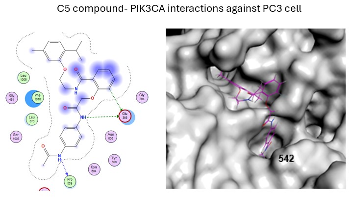
Abstract. Advances to develop new anticancer drug continue, and one of the new therapeutic targets to reach is the PI3K, due to its pathway’s functions which are related to prognosis in a variety of tumors and this protein is strongly related to tumor proliferation, migration, and apoptosis by their functions on PI3K pathway. This study evaluated a potential PI3K ligand, the C5 compound, that previously our research group proposed as a compound directed to interact with the PIK3CA´s activation region to develop a drug to regulate the PI3K´s functions. The aim of this research is to evaluate the effects of the C5 compound on PI3K´s functions related to a kind of prostate cancer without androgen receptor (PC3 cell), by in vitro cultures.We determined a regulation on the PI3K pathway in PC3 cell, due to a regulation in the expression of p-FAK, p-P70S6K and Caspase-3, these proteins were correlated to decrease the viability of the cultures to regulate the apoptosis, and this determined by western blot analysis. Then, the C5 compound was analyzed by molecular dynamics to describe the interactions of C5 compound with the PIK3CA´s activation region.We propose that the C5 compound has a high probability to be selective against PI3K, with an apoptosis (regulating Caspase-3) and cytotoxic effect on prostate cancer lines (in vitro). We also describe the possible specific interactions in the PI3K´s activation region, related to decreasing the signaling of the PI3K pathway, and probably safe for humans. These results show the high potential of this compound as a new drug against this kind of prostate cancer.
Resumen. Continúan los avances en el desarrollo de nuevos fármacos anticancerígenos, y una de las nuevas dianas terapéuticas a alcanzar es la PI3K, debido a las funciones de su vía de señalización, que están relacionadas con el pronóstico en diversos tumores. Esta proteína está fuertemente vinculada a la proliferación, migración y apoptosis tumoral a través de sus funciones en la vía de la PI3K. Este estudio evaluó un ligando potencial de la PI3K, el compuesto C5, que nuestro grupo de investigación propuso previamente como un compuesto dirigido a interactuar con la región de activación de PIK3CA para desarrollar un fármaco que regule las funciones de la PI3K. El objetivo de esta investigación es evaluar los efectos del compuesto C5 sobre las funciones de la PI3K relacionadas con un tipo de cáncer de próstata sin receptor de andrógenos (célula PC3), mediante cultivos in vitro. Determinamos una regulación de la vía PI3K en células PC3, debido a la regulación de la expresión de p-FAK, p-P70S6K y Caspasa-3. Estas proteínas se correlacionaron con la disminución de la viabilidad de los cultivos para regular la apoptosis, lo cual se determinó mediante análisis de Western blot. Posteriormente, se analizó el compuesto C5 mediante dinámica molecular para describir sus interacciones con la región de activación de PI3K.
Proponemos que el compuesto C5 tiene una alta probabilidad de ser selectivo contra PI3K, con un efecto apoptótico (regulando la Caspasa-3) y citotóxico en líneas celulares de cáncer de próstata (in vitro). También describimos las posibles interacciones específicas en la región de activación de PI3K, relacionadas con la disminución de la señalización de la vía PI3K, y probablemente seguras para humanos. Estos resultados demuestran el alto potencial de este compuesto como un nuevo fármaco contra este tipo de cáncer de próstata.
Downloads
References
. Pungsrinont, T.; Kallenbach, J.; Baniahmad, A. Int. J. Mol. Sci. 2021, 22, 11088. DOI: https://doi.org/10.3390/ijms222011088
2. Chandrasekar, T.; Yang, J.C.; Gao, A.C.; Evans, C.P. BMC Med. 2015, 13, 206. DOI: https://doi.org/10.1186/s12916-015-0457-6
3. He, Y.; Sun, M.M.; Zhang, G.G.; Yang, J.; Chen, K.S.; Xu, W.W.; Li, B. Signal Transduct. Target. Ther. 2021, 6, 425. DOI: https://doi.org/10.1038/s41392-021-00828-5
4. Meng, Q.; Xia, C.; Fang, J.; Rojanasakul, Y.; Jiang, B.-H. Cell. Signal. 2006, 18, 2262–2271. DOI: https://doi.org/10.1016/j.cellsig.2006.05.019
5. Liu, R.; Chen, Y.; Liu, G.; Li, C.; Song, Y.; Cao, Z.; Li, W.; Hu, J.; Lu, C.; Liu, Y. Cell Death Dis. 2020, 11, 797. DOI: https://doi.org/10.1038/s41419-020-02998-6
6. Shorning, B.Y.; Dass, M.S.; Smalley, M.J.; Pearson, H.B. Int. J. Mol. Sci. 2020, 21, 4507. DOI: https://doi.org/10.3390/ijms21124507
7. Glaviano, A.; Foo, A.S.C.; Lam, H.Y.; Yap, K.C.H.; Jacot, W.; Jones, R.H.; Eng, H.; Nair, M.G.; Makvandi, P.; Geoerger, B.; et al. Mol. Cancer 2023, 22, 138. DOI: https://doi.org/10.1186/s12943-023-01827-6
8. Vique‐Sánchez, J.L.; Benítez‐Cardoza, C.G. ChemistrySelect. 2022, 7. DOI: https://doi.org/10.1002/slct.202202301
9. Vanwelkenhuyzen, J.; Van Bos, E.; Van Bruwaene, S.; Lesage, K.; Maes, A.; Üstmert, S.; Lavent, F.; Beels, L.; Grönberg, H.; Ost, P.; et al. Eur. Urol. Open Sci. 2023, 53, 63–66. DOI: https://doi.org/10.1016/j.euros.2023.05.008
10. Pedrani, M.; Barizzi, J.; Salfi, G.; Nepote, A.; Testi, I.; Merler, S.; Castelo-Branco, L.; Mestre, R.P.; Turco, F.; Tortola, L.; et al. Int. J. Mol. Sci. 2025, 26, 318. DOI: https://doi.org/10.3390/ijms26010318
11. Rodriguez-Laguna, L.; Ibañez, K.; Gordo, G.; Garcia-Minaur, S.; Santos-Simarro, F.; Agra, N.; Vallespín, E.; Fernández-Montaño, V.E.; Martín-Arenas, R.; del Pozo, Á.; et al. Genet. Med. 2018, 20, 882–889. DOI: https://doi.org/10.1038/gim.2017.200
12. González-Billalabeitia, E.; Seitzer, N.; Song, S.J.; Song, M.S.; Patnaik, A.; Liu, X.-S.; Epping, M.T.; Papa, A.; Hobbs, R.M.; Chen, M.; et al. Cancer Discov. 2014, 4, 896–904. DOI: https://doi.org/10.1158/2159-8290.CD-13-0230
13. Netland, I.A.; Førde, H.E.; Sleire, L.; Leiss, L.; Rahman, M.A.; Skeie, B.S.; Miletic, H.; Enger, P.Ø.; Goplen, D. J. Neurooncol. 2016, 129, 57–66. DOI: https://doi.org/10.1007/s11060-016-2158-1
14. Akinleye, A.; Avvaru, P.; Furqan, M.; Song, Y.; Liu, D. Hematol. Oncol. 2013, 6, 88. DOI: https://doi.org/10.1186/1756-8722-6-88
15. Pomella, S.; Cassandri, M.; Braghini, M.R.; Marampon, F.; Alisi, A.; Rota, R. Int. J. Mol. Sci. 2022, 23, 1998. DOI: https://doi.org/10.3390/ijms23041998
16. Hu, H.-H.; Wang, S.-Q.; Shang, H.-L.; Lv, H.-F.; Chen, B.-B.; Gao, S.-G.; Chen, X.-B. Front. Pharmacol. 2024, 15. DOI: https://doi.org/10.3389/fphar.2024.1274209
17. Konstantinidou, G.; Ramadori, G.; Torti, F.; Kangasniemi, K.; Ramirez, R.E.; Cai, Y.; Behrens, C.; Dellinger, M.T.; Brekken, R.A.; Wistuba, I.I.; et al. Cancer Discov. 2013, 3, 444–457. DOI: https://doi.org/10.1158/2159-8290.CD-12-0388
18. Magnuson, B.; Ekim, B.; Fingar, D.C. Biochem. J. 2012, 441, 1–21. DOI: https://doi.org/10.1042/BJ20110892
19. Artemenko, M.; Zhong, S.S.W.; To, S.K.Y.; Wong, A.S.T. Cancer Lett. 2022, 535, 215593. DOI: https://doi.org/10.1016/j.canlet.2022.215593
20. Foster, P.; Yamaguchi, K.; Hsu, P.P.; Qian, F.; Du, X.; Wu, J.; Won, K.-A.; Yu, P.; Jaeger, C.T.; Zhang, W.; et al. Mol. Cancer Ther. 2015, 14, 931–940. DOI: https://doi.org/10.1158/1535-7163.MCT-14-0833
21. Ghotaslou, A.; Samii, A.; Boustani, H.; Kiani Ghalesardi, O.; Shahidi, M. Rep. Biochem. Mol. Biol. 2022, 11, 111–124. DOI: https://doi.org/10.52547/rbmb.11.1.111
22. ChemBridge Corp. Available online: https://chembridge.com/screening_libraries/#EXPRESSPick, accessed in January 2025
23. RCSB, Protein Data Bank. Available online: https://www.rcsb.org/, accessed in January 2025.
24. Mosmann, T. J. Immunol. Methods 1983, 65, 55–63.
25. Eisenbrand, G.; Pool-Zobel, B.; Baker, V.; Balls, M.; Blaauboer, B.J.; Boobis, A.; Carere, A.; Kevekordes, S.; Lhuguenot, J.-C.; Pieters, R.; et al. Food Chem. Toxicol. 2002, 40, 193–236. DOI: https://doi.org/10.1016/S0278-6915(01)00118-1
26. Wang, L.; Wang, L.; Wang, R.; Xu, T.; Wang, J.; Cui, Z.; Cheng, F.; Wang, W.; Yang, X. Heliyon 2024, 10, e31639. DOI: https://doi.org/10.1016/j.heliyon.2024.e31639
27. Jelínek, M.; Balušíková, K.; Schmiedlová, M.; Němcová-Fürstová, V.; Šrámek, J.; Stančíková, J.; Zanardi, I.; Ojima, I.; Kovář, J. Cancer Cell Int. 2015, 15, 8. DOI: https://doi.org/10.1186/s12935-015-0155-7
28. Gui, C.; Zhang, C.; Xiong, X.; Huang, J.; Xi, J.; Gong, L.; Huang, B.; Zhang, X. Int. J. Oncol. 2021, 58, 409–418. DOI: https://doi.org/10.3892/ijo.2021.5172
29. Jamalzadeh, L.; Ghafoori, H.; Aghamaali, M.; Sariri, R. Iran. J. Biotechnol. 2017, 15, 157–165. DOI: https://doi.org/10.15171/ijb.1567
30. Anguiano-Sevilla, L.A.; Lugo-Cervantes, E.; Ordaz-Pichardo, C.; Rosas-Trigueros, J.L.; Jaramillo-Flores, M.E. Int. J. Mol. Sci. 2018, 19, 3765. DOI: https://doi.org/10.3390/ijms19123765
31. Balvan, J.; Krizova, A.; Gumulec, J.; Raudenska, M.; Sladek, Z.; Sedlackova, M.; Babula, P.; Sztalmachova, M.; Kizek, R.; Chmelik, R.; et al. PLoS One. 2015, 10, e0121674. DOI: https://doi.org/10.1371/journal.pone.0121674
32. Guvenir Celik, E.; Eroglu, O. Mol. Biol. Rep. 2023, 50, 319–329. DOI: https://doi.org/10.1007/s11033-022-08034-4
33. Mirsanei, J.S.; Nazari, M.; Shabani, R.; Govahi, A.; Eghbali, S.; Ajdary, M.; Mehdizadeh, R.; Mousavi, A.S.; Mehdizadeh, M. Rep. Biochem. Mol. Biol. 2023, 12, 233–240. DOI: https://doi.org/10.61186/rbmb.12.2.233
34. Cookson, M.S.; Roth, B.J.; Dahm, P.; Engstrom, C.; Freedland, S.J.; Hussain, M.; Lin, D.W.; Lowrance, W.T.; Murad, M.H.; Oh, W.K.; et al. J. Urol. 2013, 190, 429–438. DOI: https://doi.org/10.1016/j.juro.2013.05.005
35. Jernberg, E.; Bergh, A.; Wikström, P. Endocr. Connect. 2017, 6, R146–R161. DOI: https://doi.org/10.1530/EC-17-0118
36. Chandrasekar, T.; Yang, J.C.; Gao, A.C.; Evans, C.P. Transl. Androl. Urol. 2015, 4, 365–380. DOI: https://doi.org/10.3978/j.issn.2223-4683.2015.05.02
37. Hassan, A.; Aubel, C. Cancers (Basel). 2025, 17, 2232. DOI: https://doi.org/10.3390/cancers17132232
38. Sen, A.; Khan, S.; Rossetti, S.; Broege, A.; MacNeil, I.; DeLaForest, A.; Molden, J.; Davis, L.; Iversrud, C.; Seibel, M.; et al. Mol. Oncol. 2025, 19, 225–247. DOI: https://doi.org/10.1002/1878-0261.13703
Downloads
Published
Issue
Section
License
Copyright (c) 2026 Victor Garcia-González, Ana Brenda Ortega Morales , Anna C. Navarro Padrón, Octavio Galindo-Hernández, José L. Vique-Sánchez

This work is licensed under a Creative Commons Attribution-NonCommercial 4.0 International License.
Authors who publish with this journal agree to the following terms:
- Authors retain copyright and grant the journal right of first publication with the work simultaneously licensed under a Creative Commons Attribution License that allows others to share the work with an acknowledgement of the work's authorship and initial publication in this journal.
- Authors are able to enter into separate, additional contractual arrangements for the non-exclusive distribution of the journal's published version of the work (e.g., post it to an institutional repository or publish it in a book), with an acknowledgement of its initial publication in this journal.